Description
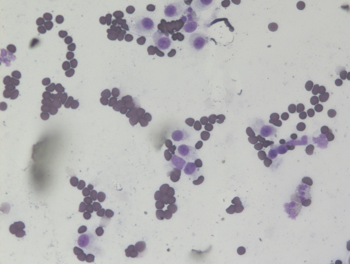
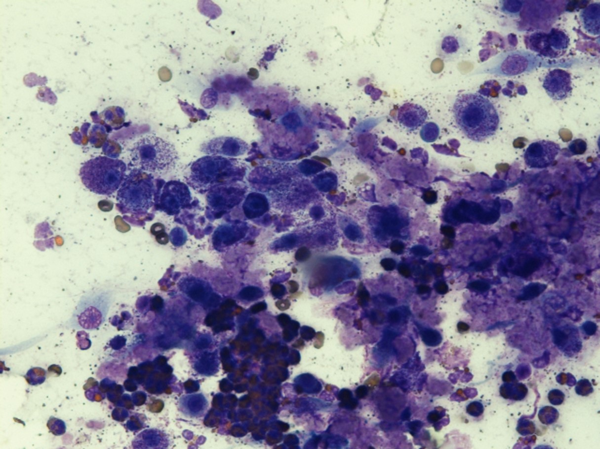
A mast cell tumour consists of cancerous mast cells. A mast cell is a certain type of white blood cell that would normally protect the body against parasites (viruses and bacteria are dealt with by other white blood cells) but can also contribute to allergic reactions.
Therefore, these cells can mainly be found in tissues adjacent to the outside world, such as the skin, the respiratory and digestive system.
Behaviour
A remarkable aspect of mast cells is that they contain a large number of granules, which in turn contain inflammatory substances (such as histamine, heparin, prostaglandins, platelet activating factors, leukotrienes, substances attracting other white blood cells such as neutrophils and eosinophils, enzymes that degrade proteins, serotonin). When a mast cell encounters a parasite, it will release the contents of its granules to damage that parasite and cause a local inflammation.
This characteristic is of importance when dealing with cancerous mast cells. Some cancerous mast cells contain a lot of granules which can release their contents at the slightest stimulus. This release can lead to local inflammation, itching, digestive ulcers, and in severe cases to a serious drop in blood pressure or shock and other symptoms associated with serious allergic reactions.
Most mast cell tumors metastasize first towards the regional lymph nodes, afterwards mainly towards the liver (46%) and the spleen (41%). Other organs can be involved as well, lung metastases are very rare.
Prevalence
Mast cell tumours are the most frequent skin tumour in dogs (16-21%) and are mainly seen in middle-aged dogs.
Breed predisposition
The following dog breeds are more prone to develop this tumour type than other breeds: Beagle, Boston Terrier, Boxer, Bulldog, Cocker Spaniel, Golden retriever, Labrador retriever, Pug, Rhodesian Ridgeback, Schnauzer, Shar-Pei, Scottish Terrier, Staffordshire Bull Terrier and Weimaraner. Differences have been reported between breeds, e.g. Boxers usually experience a more benign form than Shar-Peis.
Symptoms
Mostly, mast cell tumours are well delineated and appear as a single lump. In 11-14% of cases more than one tumour will be present.
If a mast cell tumour is well differentiated (= if a clearly recognizable organization is present within the tumour), it will manifest itself as a single lump, have a size between 1 to 4 cm, grow slowly, rarely develop ulcers and can be present for years without causing any issues.
If a mast cell tumour is poorly differentiated (= if there is no clearly recognizable organization present within the tumour), it tends to grow rather fast, potentially become very large, develop ulcers and lead to local irritation. The surrounding tissues are often swollen, and satellite lumps may develop.
When a mast cell tumour is stimulated (e.g. manipulation by owner/veterinarian, trauma (e.g. when the dog gnaws on it), radiation, treatment with heat/cold), the mast cells can release inflammatory substances. As a result, the tumour will swell and a red ring will become visible around the tumour. The latter is called the sign of Darier. The swelling and redness will disappear by itself after a while, if the dog does not continue to scratch or lick.
The larger the tumour and/or the more tumours there are, the more dangerous the effects of the inflammatory reaction can become. The inflammatory substances can cause many side effects, such as itching, digestive ulcers (associated symptoms are vomiting (blood), decreased appetite, diarrhea, black feces and stomachache), clotting anomalies, low blood pressure and shock. Gastric ulcers occur as a consequence of blood vessel damage, an increase in gastric acid and an overly contracting stomach. On autopsy or during surgery, a gastric or intestinal ulcer is found in 35-83% of dogs with a mast cell tumour.